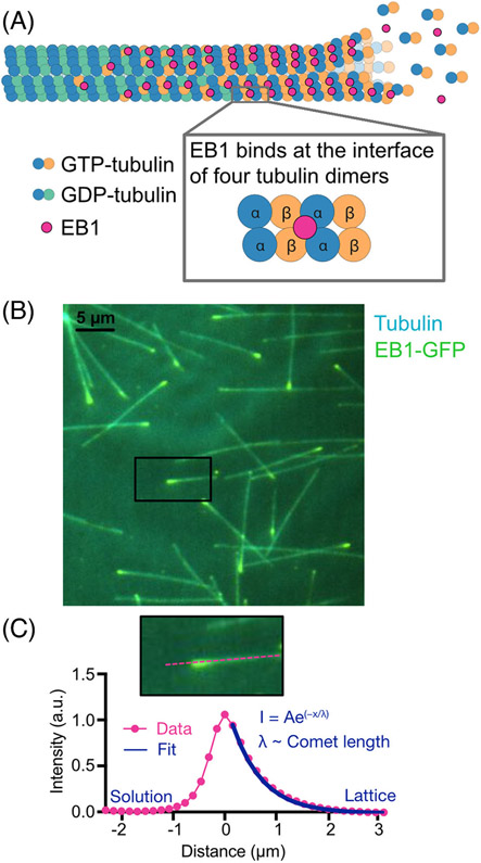
FIGURE 2.
EB localization on growing microtubule ends serves as a proxy for the GTP-cap. (A) EBs bind at the interface of four tubulin dimers, in a location that facilitates EB’s recognition of the nucleotide state of tubulin within the microtubule lattice. Due to its nucleotide sensitivity, EB localization can be used to mark the GTP-cap. (B) Total-internal-reflection-fluorescence image of EB1-GFP localization on ends of growing microtubules in vitro. (C) The fluorescent EB1-GFP signal resembles a comet-like shape, exponentially decaying along the microtubule lattice. Fluorescent comet profiles are averaged together then fit to an exponential decay function and the decay constant is used as a proxy for the length of the GTP-cap.

