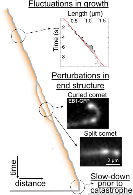
FIGURE 4.
Light microscopy provides hints into dynamic changes at growing microtubule ends. A time–distance schematic (kymograph) of a growing microtubule end. (Top) Microtubule ends can exhibit significant fluctuations in end position over time.[83,84] (©2021 Farmer et al. Inset originally published in the Journal of Cell Biology. https://doi.org/10.1083/jcb.202012144) (Middle) Observations of growing microtubule ends reveal occurrences of microtubule end splitting, curling and repair.[41,73,123] (Bottom) Microtubules typically exhibit a slow-down in growth accompanied by the loss of the EB-comet prior to catastrophe.[34,41,124]

