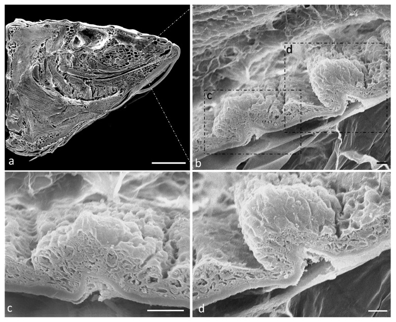
Figure 1.
(a) Scanning electron microscope micrograph of a sagittal section of a zebrafish head. (b) Scanning electron microscope of two taste buds with different shapes. Higher magnification of onion-shaped taste bud (c) and pear-shaped taste bud (d). Scale bar: 1 mm (a) 10 μm (b) 20 μm (c) 10 μm (d).

