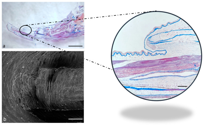
Figure 4.
(a) Stereomicrograph of a sagittal section of the tongue (Masson trichrome with aniline blue staining): apex of the tongue (circular insert). (b) Scanning electron microscope micrograph of the tongue apex. Magnification 4×, scale bar 1 mm (a). Scale bar: 1 mm (b). Magnification 10×, scale bar 200 µm (insert).

