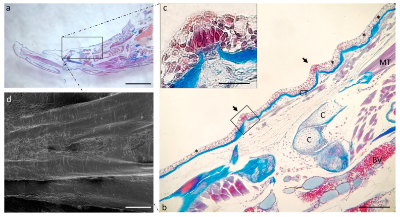
Figure 5.
(a) Stereomicrograph of a sagittal section of the tongue: dorsal surface of the tongue body (insert). (b) Light micrographs of the dorsal surface of the tongue with taste buds (arrows). Connective tissue (CT); cartilage (C); muscular tissue (MT); blood vessel (BV); goblet cells (asterisks). (c) high magnification of taste bud in image. (b) Masson trichrome with aniline blue staining. (d) Scanning electron microscope micrograph of the tongue body. (a) Magnification 4×, scale bar 1 mm. (b) Magnification 10×, scale bar 200 µm. (c) Magnification 63× oil, scale bar 10 µm. (d) Scale bar: 1 mm.

