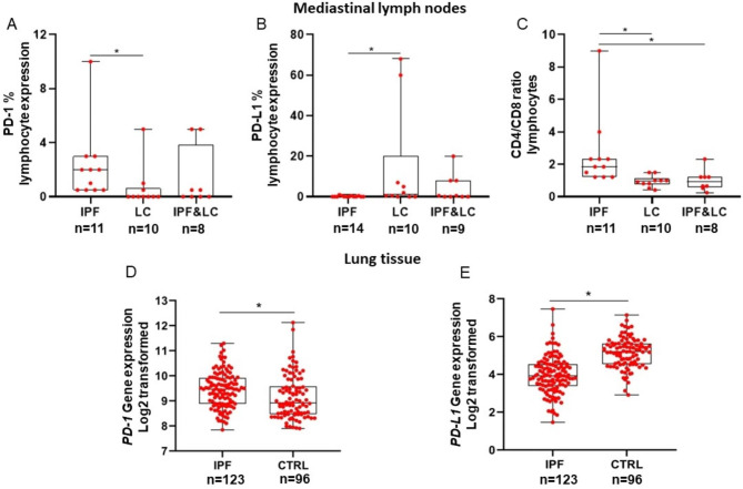
Fig. 2.
Mediastinal lymph nodes: Median PD-1% expression was significantly higher in lymphocytes of patients with IPF compared to lung cancer (IPF: 2.0, 95% CI: 0.5 to 3.0 vs. lung cancer: 0.0, 95% CI: 0.0 to 0.8, Kruskal–Wallis test; p = 0.02), (Panel A). Median PD-L1% expression was significantly lower in lymphocytes of patients with IPF compared to lung cancer (IPF: 0.0, 95% CI: 0.0 to 0.5 vs. lung cancer: 1.3, 95% CI: 0.0 to 34.8, Kruskal–Wallis test; p = 0.04), (Panel B). Median CD4/CD8 ratio was significantly higher in mediastinal lymph nodes of patients with IPF compared to patients with lung cancer and patients with concomitant IPF/lung cancer (IPF: 1.9, 95% CI: 1.2 to 2.6, vs. lung cancer: 0.9, 95% CI: 0.7 to 1.3, vs. IPF and lung cancer: 0.9, 95% CI: 0.5 to 1.4, Kruskal–Wallis test; p = 0.001), (Panel C). Lung tissue: Analysis of the Lung Genomics Research Consortium cohort showed significantly increased expression of PD-1 and decreased expression of PD-L1 in patients with IPF (n = 123) compared to control (CTRL) subjects with normal lung histology (n = 96), (PD-1: IPF: 9.5, 95% CI: 9.3 to 9.6, vs. controls: 8.9, 95% CI: 8.8 to 9.3, Mann- Whitney test; p = 0.001), (PD-L1: IPF: 4.0 ± 1 vs. controls: 5.1 ± 0.8, Unpaired t test; p < 0.0001) (Panel D and E)

