
Figure 3.
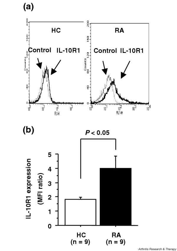
(a) Cell surface expression of type 1 interleukin-10 receptor (IL-10R1) on CD4+ T cells from patients with rheumatoid arthritis (RA) and from healthy controls (HC). Peripheral blood mononuclear cells were stained with anti-IL-10R1 antibody or with isotype-matched control antibody, followed by incubation with FITC-conjugated goat anti-mouse IgG1 polyclonal antibody, and were then stained with phycoerythrin-conjugated anti-CD4 mAb. The expression of CD4 and IL-10R1 was determined by flow cytometric analysis. Representative histographic patterns of IL-10R1 expression on CD4+ T cells from RA patients and HC are shown. (b) The intensity of IL-10R1 on CD4+ T cells was expressed as the ratio of the mean fluorescence intensity (MFI) of staining with anti-IL-10R1 to control antibody. Values are the mean ± standard error of the mean. n, number of samples tested.