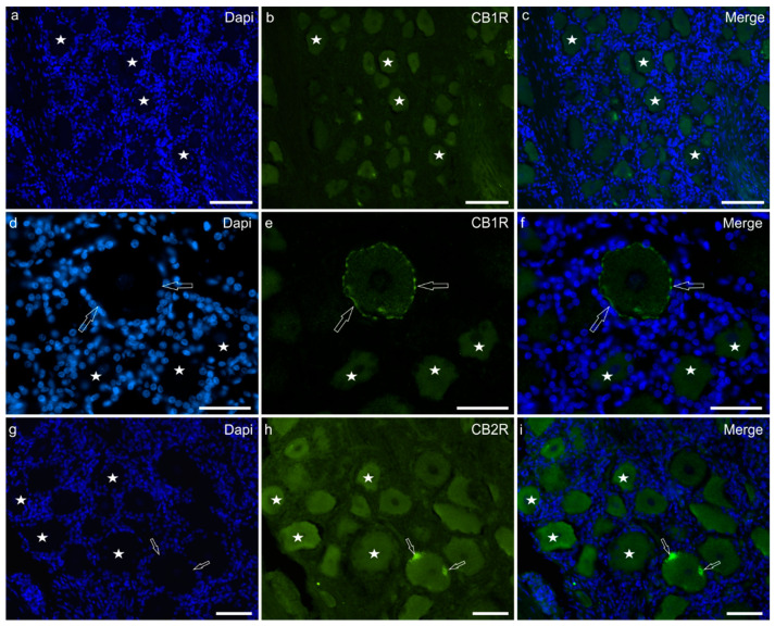
Figure 1.
Photomicrographs of equine trigeminal ganglion cryosections showing immunoreactivity for the cannabinoid receptor type 1 (CB1R) (a–f) and type 2 (CB2R) (g–i). (a–f) Stars indicate a few sensory neurons showing faint cytoplasmic CB1R immunoreactivity of the cell body cytoplasm. The open arrows indicate a neuron in which CB1R immunoreactivity was also expressed by the cell membrane. (g–i) Stars indicate neurons expressing moderate CB2R immunoreactivity. Two open arrows indicate autofluorescent pigments which were confined to the edges of the cell. Bar: 50 µm.

