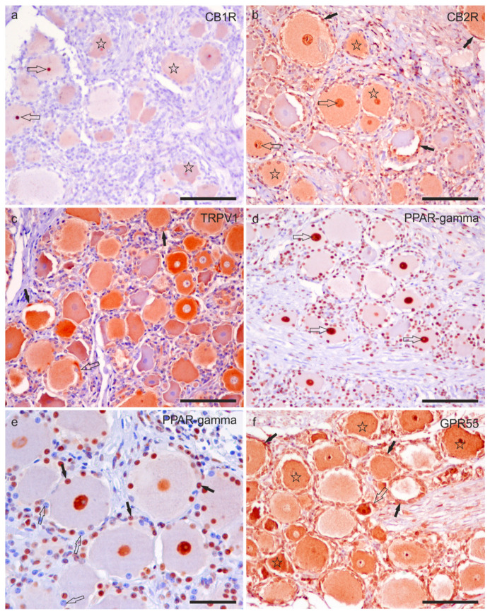
Figure 4.
Photomicrographs of formalin-fixed paraffin embedded sections of equine trigeminal ganglion showing immunoreactivity for the cannabinoid receptor type 1 (CB1R) (a) and type 2 (CB2R) (b), transient receptor potential vanilloid 1 (TRPV1) (c), perossisome proliferator-activated receptor gamma (PPARγ) (d,e), and G-protein coupled receptor 55 (GPR55) (f). (a) Stars indicate sensory neurons showing faint CB1R immunoreactivity. The open arrows indicate two strongly immunoreactive nucleoli for the CB1R. (b) Stars indicate sensory neurons which showed moderate CB2R immunoreactivity. The open arrows indicate the neuronal nuclei expressing strong CB2R immunoreactivity. Black arrows show satellite glial cells surrounding the sensory neurons, showing faint-to-moderate CB2R immunoreactivity. (c) A large proportion of sensory neurons expressed moderate-to-strong TRPV1 immunoreactivity. Axon hillocks (open arrow) were also positive for TRPV1, as well as satellite glial cells (black arrows). (d) The open arrows indicate the nuclei of the sensory neurons, showing strong PPARγ immunoreactivity. (e) Not all nuclei of satellite glial cells showed PPARγ immunoreactivity; some were strongly reactive (black arrows), whereas some appeared negative (open arrows). (f) Stars indicate the cell body cytoplasm of sensory neurons with moderate-to-strong GPR55 immunoreactivity. The open arrows indicate the axon hillock of a small neuron which expressed strong GPR55 immunoreactivity. The black arrows indicate some satellite glial cells which were positive for GPR55. Bars: (a–d,f): 100 µm; (e): 50 µm.

