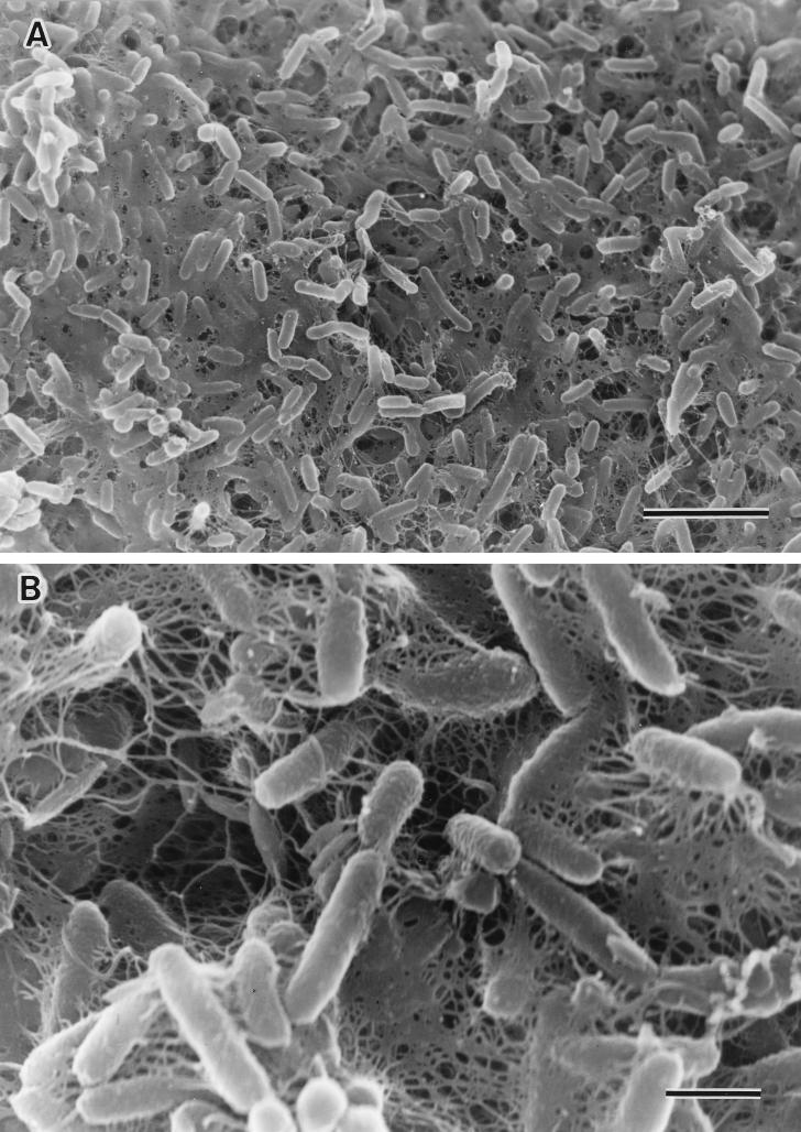
FIG. 5.
Scanning electron micrographs of biofilm formation by V. cholerae O1 strain TSI-4/R. (A) Most of the surface has been colonized with actively dividing rod cells, and finger-like projections of extracellular polymeric material are present. Bar, 5 μm. (B) High magnification indicates the presence of extracellular polymeric materials on the surfaces of bacterial cells. Bar, 1 μm.

