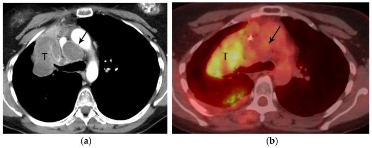
Figure 7.
N2 nodal metastasis. (a) CT shows a 6.5 cm tumor (T) in the right upper lobe and low attenuation right paratracheal adenopathy (arrow). (b) PET/CT shows FDG avidity of the primary tumor (T) but the biopsy-proven N2 ipsilateral nodal metastasis is not FDG avid. Necrotic nodal metastases can give false negative results on PET/CT.

