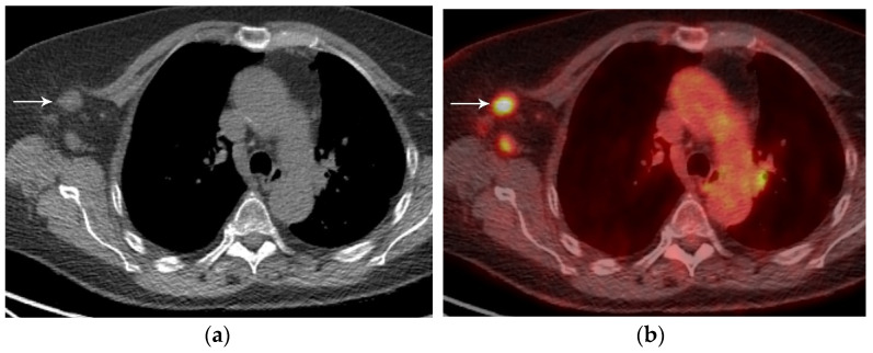
Figure 9.
Left lung cancer and right axillary nodal metastases. (a) CT, and (b) axial PET/CT show FDG-avid right axillary adenopathy (arrow). Biopsy confirmed metastatic disease from the left lung cancer. The N status represents regional spread of disease. Lymph nodes not addressed in N classification, such as internal mammary, axillary, and retroperitoneal, represent distant metastatic disease.

