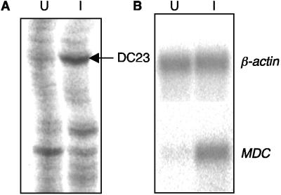
FIG. 2.
Up-regulation of MDC in DCs during S. enterica serovar Typhimurium infection. (A) Differential display was used to identify differentially expressed genes in DCs following S. enterica serovar Typhimurium BRD509/GF3 infection. A portion of a differential display gel is shown. The arrow indicates the position of a differentially displayed band whose intensity was reduced in uninfected DCs (lane U) compared with infected DCs (lane I). Subsequent sequencing and a BLAST search against the GenBank database assigned this differential display product to the MDC gene. (B) Northern blotting was used to confirm the increased expression of MDC. RNA was extracted from sorted uninfected and infected DCs, hybridized with the MDC-specific DNA probe, and then washed and rehybridized with the β-actin gene probe. The hybridization pattern was analyzed with a phosphorimager.

