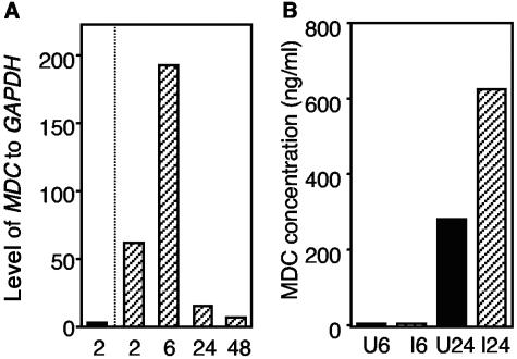
FIG. 3.
MDC expression in DCs. (A) Real-time RT-PCR was used to study the kinetic expression of the MDC gene in DCs following S. enterica serovar Typhimurium infection and sorting of infected and uninfected cells. The data are the relative expression levels of MDC compared to the expression levels of GAPDH in uninfected DCs which were sorted 2 h after mock infection (solid bar) or in S. enterica serovar Typhimurium-infected DCs sorted at different times postinfection (in hours) (cross-hatched bars). The results of one of two experiments are shown, and the results were similar in the different experiments. (B) MDC protein expression as determined by an ELISA (detection limit, <0.04 ng/ml). At 6 and 24 h after infection and sorting, culture supernatants from uninfected DCs (solid bars) and infected DCs (cross-hatched bars) were collected, and the amounts of MDC in the supernatants were analyzed. U6 and U24, uninfected cells at 6 and 24 h postinfection, respectively; I6 and I24, infected cells at 6 and 24 h postinfection, respectively.

