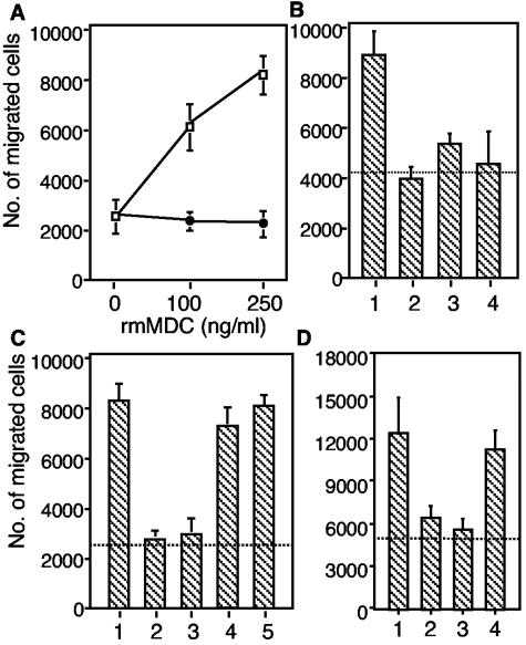
FIG. 4.
MDC-induced chemotaxis of CD4 + T cells. Purified CD4 + cells (1.5 × 10 5 cells) were added to the upper wells of a microchamber, and medium was added to the lower chamber. The data are the means and standard errors for duplicate samples. (A) Migratory responsiveness of CD4 + T cells to increasing doses of rmMDC (□) and in the presence of 10 μg of rat anti-mouse MDC antibody per ml (•). (B) Migration of CD4 + T cells induced by MDC in the DC culture supernatant. Bar 1, infected DC supernatant diluted 1:5; bar 2, infected DC supernatant diluted 1:5 plus anti-mouse MDC; bar 3, uninfected DC supernatant diluted 1:5; bar 4, uninfected DC supernatant diluted 1:5 plus anti-mouse MDC. (C) Migration of CD4 + T cells was inhibited by rabbit anti-MDC antiserum but not by the control antiserum. Bar 1, 250 ng of rmMDC per ml; bar 2, rmMDC plus anti-MDC antiserum diluted 1:5; bar 3, rmMDC plus anti-MDC antiserum diluted 1:50; bar 4, rmMDC plus control antiserum diluted 1:5; bar lane 5, rmMDC plus control antiserum diluted 1:50. (D) Bar 1, infected DC supernatant diluted 1:5; bar 2, infected DC supernatant diluted 1:5 plus anti-mouse MDC; bar 3, infected DC supernatant diluted 1:5 plus anti-MDC antiserum diluted 1:10; bar 4, infected DC supernatant diluted 1:5 plus control anti-GST antiserum diluted 1:10. The dotted lines indicate the background migration observed in medium-only control samples.

