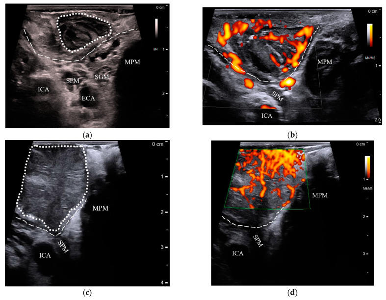
Figure 2.
Comparison of transoral US features in benign and cancerous palatine tonsils: (a) a normal palatine tonsil (dotted outline) with striated crypts overlying the constrictor muscle (dashed line), lying superficially to the stylopharyngeus muscle (SPM), styloglossus muscle (SGM), medial pterygoid muscle (MPM), internal carotid artery (ICA), and external carotid artery (ECA); (b) power doppler shows the palatine tonsils’ blood flow originating from the constrictor muscle and branching outward parallel to the crypts; (c) a stage-T2 palatine tonsil HPV+ SCC appears as an enlarged, hypoechoic mass without crypt striations (dotted outline), but respects the boundaries of the constrictor muscle (dashed line); (d) with power doppler, the same tumor is seen with random, chaotically increased blood flow. US: ultrasound; HPV+ SCC: human papillomavirus-positive squamous cell carcinoma.

