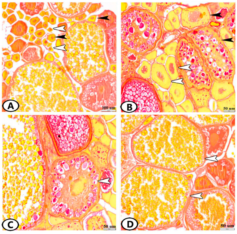
Figure 1.
Histological structure of the ovary of zebrafish stained by Sirus red. (A) Oogonia (white arrowheads) and chromatin nucleolus stage (black arrowheads). (B) Perinucleolar stage (white arrowheads) and vacuolated follicles (black arrowheads). Note that the nucleoplasm attained red granules with Sirus red stain. (C) Yolk globule stage (white arrowhead). (D) Mature stage (white arrowheads).

