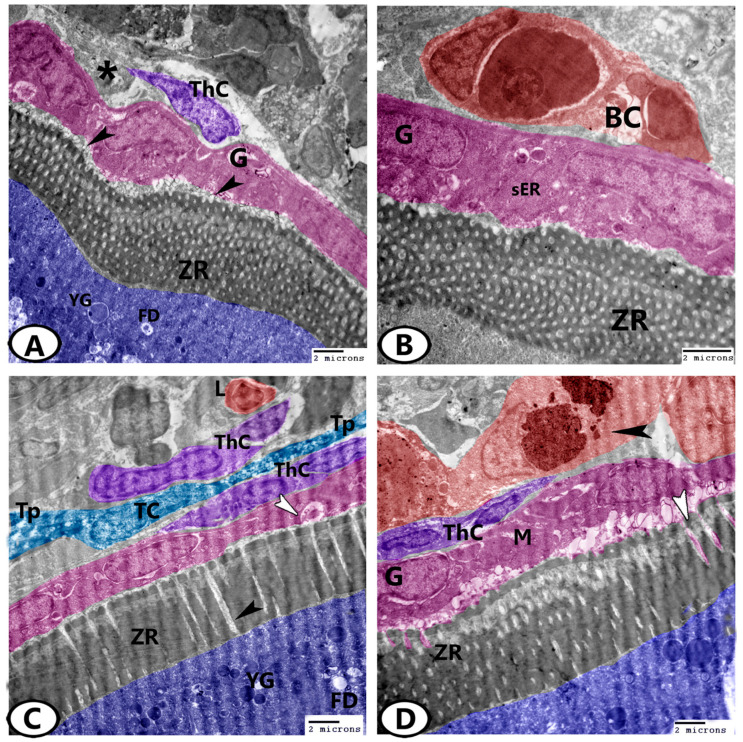
Figure 6.
Digital colored TEM images of the mature ovarian follicles of zebrafish. (A) Fat droplets (FD) and yolk globules (YG) are found in the outer ooplasm of the mature follicles. Note the processes (arrowheads) of the granulosa cells (G, pink) that penetrated the zona radiata (ZR). Theca cells (ThC, violet) are embedded in the collagen network (asterisk). (B) Higher magnification shows blood capillaries (BC, red) in the thecal layer. Note the granulosa (G, pink) that contained sER and zona radiata (ZR). (C) The zona radiata (ZR) was traversed perpendicularly by pore canals (black arrowhead) containing processes from both oocytes (blue) and zona granulosa (G, pink, white arrowhead). The oocytes contained fat droplets (FD) and yolk globules (YG). Note the presence of telocytes (TC) and telopodes (Tp) between the thecal layers (ThC). Lymphocytes (L, red) were found in the ovarian stoma. (D) The granulosa layer (G, pink) contained many mitochondria (M). Note the processes (white arrowhead) of the granulosa layer that penetrated the zona radiata (ZR). Large macrophages (red, black arrowhead) could be seen neighboring the thecal layer (ThC, violet).

