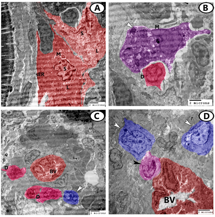
Figure 7.
Digital colored TEM images of the ovarian stroma. (A) Two steroid interstitial stromal cells (S, red) contained sER, mitochondria (M), and many lipid droplets (L). (B) Dendritic cells (D, pink) could also be seen in the ovarian stroma in association with macrophages (M, violet) that contained heterogeneous materials (arrowhead). (C) Dendritic cells (D, pink) were distributed around the blood vessels (BV). Note the presence of stem cells (arrowhead, blue) with dividing nuclei. (D) Monocytes (black arrowhead) were in close contact with blood vessels (BV). Stem cells (white arrowhead, blue) were observed in the stroma.

