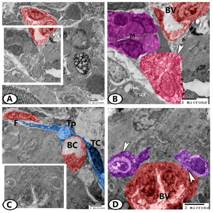
Figure 8.
Digital colored TEM images of the ovarian stroma. (A,B) Low and higher magnifications show the fat cells (pink, arrowhead) and macrophages (violet, M) in association with blood vessels (BV, red). (C) Telocytes (TC, blue) with telopodes (Tp) contained secretory vesicles extended around the blood capillaries (BC) and contacted adjacent fibroblasts (F, red). (D) Higher magnification of the boxed area in (C) shows many endocrine cells (violet) with electron-dense granules (arrowheads) in close contact with the blood vessels (BV).

