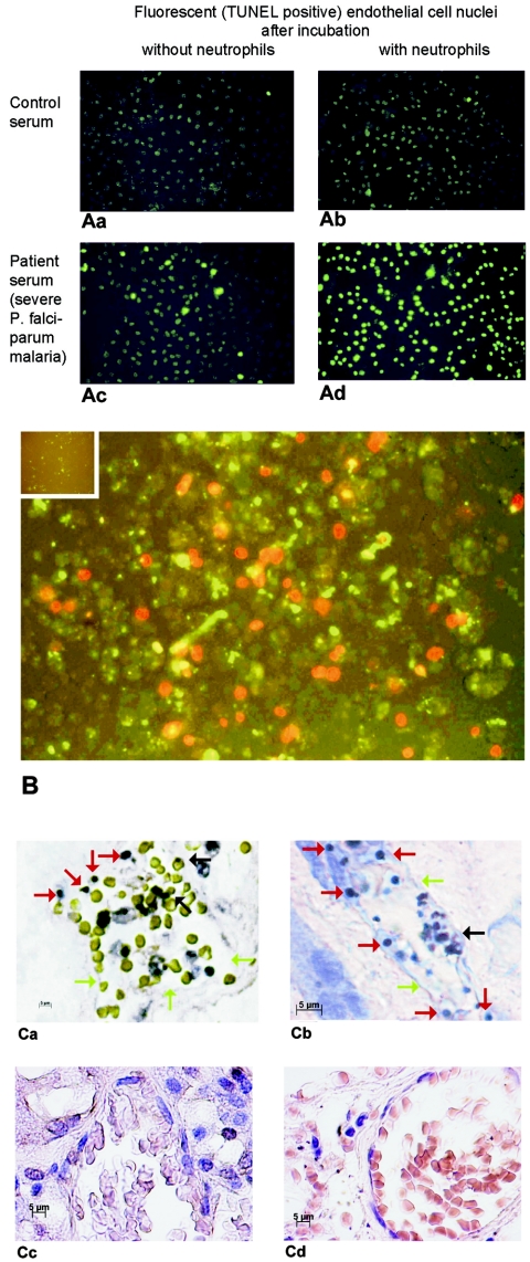
FIG. 1.
(A) TUNEL-stained nuclei from endothelial cells incubated with serum from a patient with fatal P. falciparum malaria (Table 1, patient 2) and from a healthy control, with or without neutrophils. (B) Annexin V-stained endothelial cells incubated with neutrophils plus serum from a patient with fatal P. falciparum malaria (green fluorescence). Necrotic cells were stained with propidium iodide (red fluorescence). The inset shows endothelial cells incubated with serum from a healthy person as a negative control. (C) TUNEL-stained kidney (a) and lung (b) sections from two patients who had died from P. falciparum malaria. The capillaries contain erythrocytes and malaria pigment (black arrows). Much of the endothelial cell lining is missing (green arrows). The endothelial cell nuclei are round, condensed, and TUNEL positive, indicating apoptosis (red arrows). Normal capillary endothelial cells in kidney (c) and lung (d) sections from a traffic accident victim are also shown.

