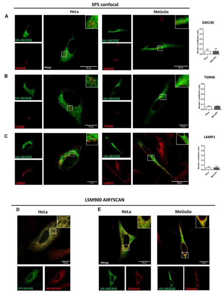
Figure 4.
Subcellular localization of HA-ABCB5β in transfected HeLa and MelJuSo cells. (A–C) A total of 48 h post-transfection with HA-ABCB5β, HeLa, or MelJuSo cells were fixed with 4% of paraformaldehyde and processed for the detection of HA-ABCB5β (green) and different organelle markers (red): (A) GM130 for the cis-Golgi apparatus, (B) TGN46 for the trans-Golgi apparatus, (C) LAMP1 for late endosomes and lysosomes. Micrographs were obtained using an SP5 confocal microscope. Graphs show the quantification of co-localization extent by using Mander’s coefficient. n = 10 cells were analyzed from at least 3 independent experiments. (D) Analysis of co-localization between GFP-ABCB5β (green) and HA-ABCB5β (red, detected using an anti-HA antibody) with a Zeiss Airyscan microscope, 48 h after co-transfection in HeLa cells. (E) Higher resolution micrographs of HA-ABCB5—calnexin colocalization taken using a Zeiss LSM900 microscope. Scale bar = 25 or 10 µm. Magnified views in right insets.

