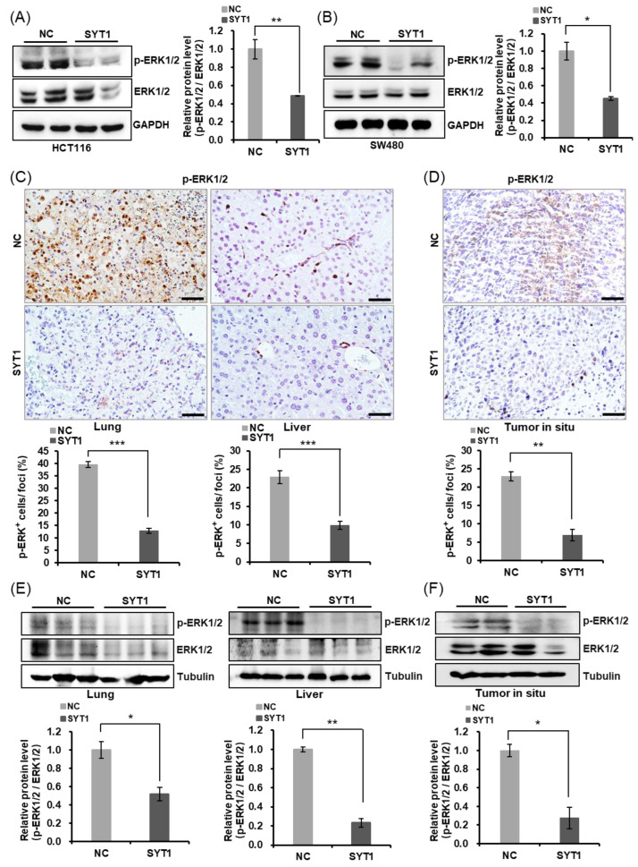
Figure 7.
SYT1 inhibited ERK/MAPK signaling. (A,B) Western blots of MAPK42/44 (ERK1/2) and phosphorylated MAPK42/44 (p-ERK1/2) in HCT116 (A) and SW480 (B) cells at control or SYT1 overexpression. (C) Immunohistochemical stains of p-ERK1/2 in liver and lung tissues from control and SYT1-overexpressing CRC metastasis mice model. Scale bar, 50 μm. (D) Immunohistochemical stains of p-ERK1/2 in in situ tumor of control and SYT1-overexpressing mice. Scale bar, 50 μm. (E) Western blots of p-ERK1/2 in liver and lung tissues of control and SYT1-overexpressing CRC metastasis mice model. (F) Western blots of p-ERK1/2 in orthotopic transplantation tumor of control and SYT1-overexpressing mice. Tubulin or GAPDH was used as the loading control; * p < 0.05, ** p < 0.01, and *** p < 0.001. The uncropped bolts are shown in Supplementary Materials Figure S2.

