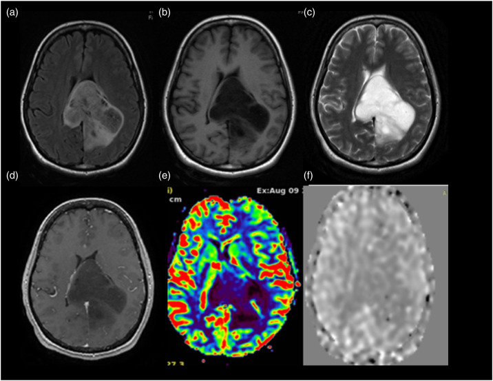
Legend 3.
Glioma WHO grade 2 IDH mutant. A 33-year-old female presented with headache of 6 months duration. MRI scan shows a T2WI (Figure (c)) hyperintense lesion in the splenium of corpus callosum with extension on either side of midline with few areas appearing hypointense on FLAIR axial (Figure (a)) suggestive of T2-FLAIR mismatch. It appears hypointense on T1WI (Figure (b)). On perfusion imaging, the lesion shows low perfusion. The value of normalized T2*DSC perfusion rCBV (Figure (e)) in the lesion is 0.76. ASL perfusion (Figure (f)) also shows low value (0.70) of normalized aCBV in the region of tumor which corresponds with the low perfusion on T2* DSC perfusion imaging. On histopathological evaluation, it turned out to be diffuse astrocytoma WHO grade 2-IDH mutant.

