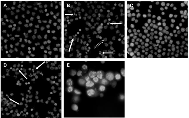
FIG. 2.
Monocyte cell death in response to C. difficile toxin A. Purified monocytes and THP-1 cells were incubated for 24 h in the absence of toxin A (A and C) or in the presence of 1,000 ng of toxin A per ml (B and D). THP-1 cells were also incubated with 30 μM etoposide for 5 h (positive control) (E). Cytospin preparations of cells were fixed and stained with 10 μg of Hoechst 33342 dye per ml before visualization with a fluorescence microscope with UV light excitation. Cells exhibiting characteristic features of apoptotic cell death are indicated by solid arrows. A number of nonapoptotic cells with diffuse nuclear staining (open arrow) were also seen after exposure to toxin A, and these cells were probably cells undergoing necrosis (see Fig. 3C).

