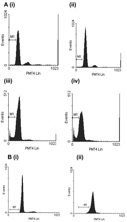
FIG. 4.
Representative DNA fluorescence profiles of propidium iodide-labeled purified preparations of control and toxin A-exposed monocytes. After incubation, the cells were fixed, permeabilized, and treated with RNase before labeling with propidium iodide and analysis of propidium iodide fluorescence. In panel A, the monocytes were incubated in the absence of C. difficile toxin A (panel i) or in the presence of 1,000 ng of C. difficile toxin A per ml for 2 h (panel ii), 5 h (panel iii), or 24 h (panel iv). In panel B, monocytes were exposed to control medium (panel i) or 30 μg of toxin A per ml (panel ii) for 5 h prior to propidium iodide labeling. Control monocytes (panels i in panels A and B) and monocytes exposed to 30 μg of toxin A per ml (panel ii in panel B) did not exhibit apoptotic characteristics, as shown by a lack of events in the hypodiploid region (M1). By contrast, many events characteristic of apoptotic cell death were seen in the hypodiploid (or sub-G1) region in monocytes incubated with 1,000 ng of toxin A per ml for 5 h (panel iii in panel A) and 24 h (panel iv in panel A).

