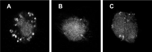
FIG. 7.
Cytochrome c expression by control and toxin A-exposed monocytes. Purified monocytes were exposed to control medium (A), 1,000 ng of toxin A per ml (B), or 30 μM etoposide (positive control) (C) for 5 h prior to fixation, permeabilization, and incubation with anti-cytochrome c antibody. Following application of FITC-conjugated goat anti-mouse antibody, the cells were examined by confocal fluorescence microscopy. Representative images are shown. Cytochrome c labeling is confined to mitochondria in panel A. By contrast, in monocytes exposed to toxin A and etoposide for 5 h (B and C), cytochrome c labeling occurs almost exclusively in the cytoplasm.

