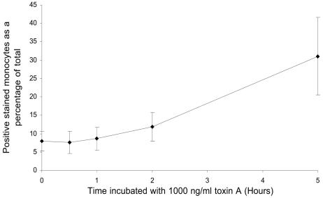
FIG. 8.
Activation of caspase-3 in monocytes exposed to C. difficile toxin A. Purified monocytes were incubated in the absence or in the presence of 1,000 ng of toxin A per ml for 0.5, 1, 2, and 5 h at 37°C prior to fixation, permeabilization, incubation with PE-conjugated anti-active caspase-3 antibody, and analysis by flow cytometry. Monocytes exhibiting positive intracellular active caspase-3 fluorescence were enumerated, and the data were expressed as a percentage of the total number of cells analyzed. The data are means ± standard errors of the means for three experiments. Compared to unstimulated cells, a significantly greater proportion of monocytes exposed to 1,000 ng of toxin A per ml for 5 h exhibited intracellular expression of activated caspase-3 (7.9% ± 2.6% versus 31.1% ± 10.5%; P < 0.05).

