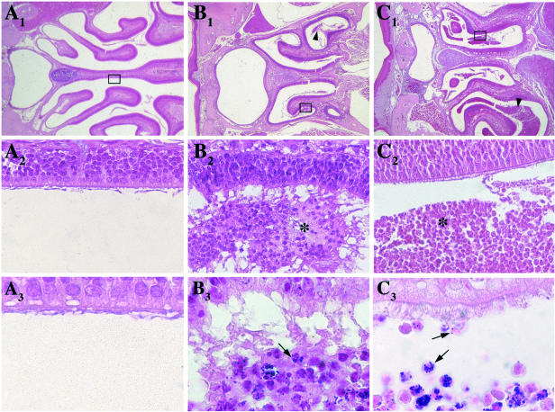
FIG. 2.
Histological sections of the nasal cavities of control BALB/c mice (A) and BALB/c and CFTR−/− mice 24 h after inoculation of strain COL (B and C, respectively). Hematoxylin-eosin (upper panel, 25× enlargement; middle panel, 400× enlargement of selected area) and Gram staining (lower panel, 1,000×) were performed with 5-μm-thick sections. Luminal cellular clumps spread along the nasal cornets are indicated with arrowheads (B1 and C1) and asterisks (B2 and C2). Bacteria that appeared to be located intracellularly within the cellular clumps composed of neutrophils and degenerated cells are denoted with an arrow (B3 and C3). In CFTR−/− mice, bacteria were present along the nasal epithelium (C3).

