
FIG. 1.
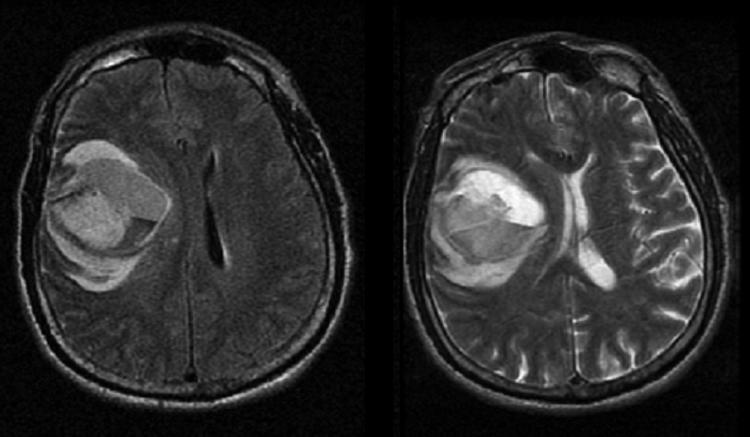
MRI of 66-year-old male after a motor vehicle accident showing a large right frontal intraparenchymal hemorrhage on the FLAIR (left image) and T2 (right image) sequences. The FLARE images shows diffuse, heterogeneous increased signal intensity consistent with evolving blood products with surrounding edema in the frontal lobe.