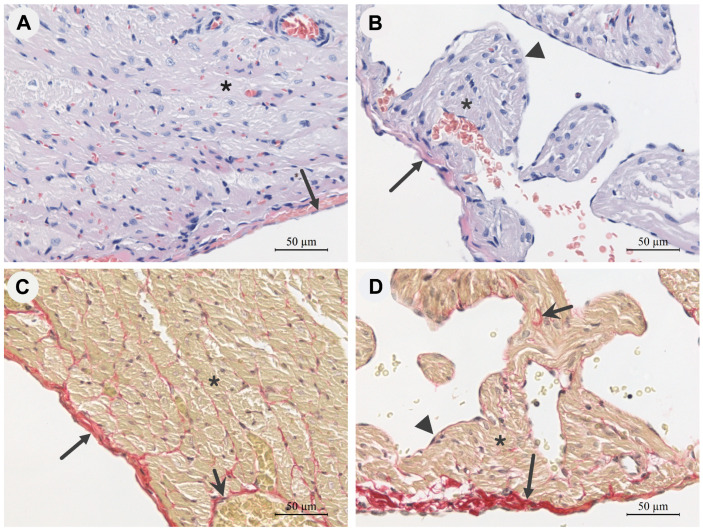
Figure 1.
Representative photomicrographs of longitudinal histological sections of the free wall of the right ventricle (A,C) and the wall of the right atrium (B,D) of guinea pig hearts under physiological conditions, objective magnification 40×. Haematoxylin and eosin staining (A,B): cell nuclei—dark purple; cytoplasm and intercellular substance—pink. Picrosirius red staining (C,D): connective tissue collagen skeleton—dark maroon fibres; cardiomyocytes—yellow-brown; cell nuclei—black. Long arrow—epicardium, short arrow—connective tissue carcass, ▲—endocardium, *—cardiomyocytes.

