Figure 1.
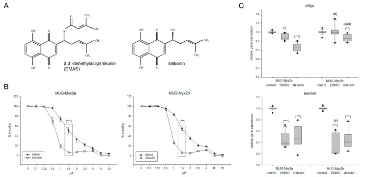
Effects of DMAS and shikonin on cell viability and proliferation. (A) Chemical structures of β,β-dimethylacrylshikonin (DMAS) and shikonin. (B) Dose–response relationship showing the dose-dependent reduction in cell growth, where shikonin showed a highly significant, more efficient effect than DMAS at a concentration of 1.5 µM. (C) Relative gene expression of the proliferation markers cMyc and survivin 24 h after treatment with the respective IC50 concentrations of DMAS/shikonin (mean ± SD; n = 6; measured in triplicate). Untreated cells were used as controls (ratio = 1). Statistical significances are defined as follows: */# p < 0.05; ** p < 0.01; ***/### p < 0.001 (controls vs. DMAS/shikonin treated cells were represented with stars; significances between both cell lines were represented with rhombuses).

