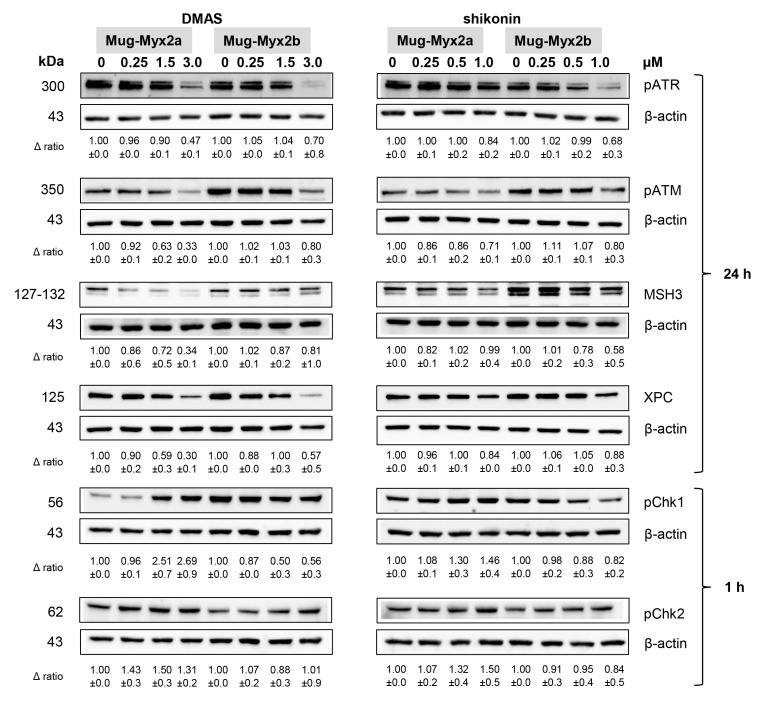
Figure 6.
DNA damage response after DMAS and shikonin treatment. Protein expression and phosphorylation of DNA damage markers pATR, pATM, MSH3, and XPC, as well as pChk1 and pChk2 was analyzed by immunoblotting under control conditions (0) and after treatment with 0.5, 1.5, and 3 µM DMAS, respectively 0.25, 0.5, and 1.0 µM shikonin. β-actin was used as loading control. Δ ratio, fold change normalized to non-treated controls (mean ± SD of n = 3). Full-length blots are presented in Supplementary Data.

