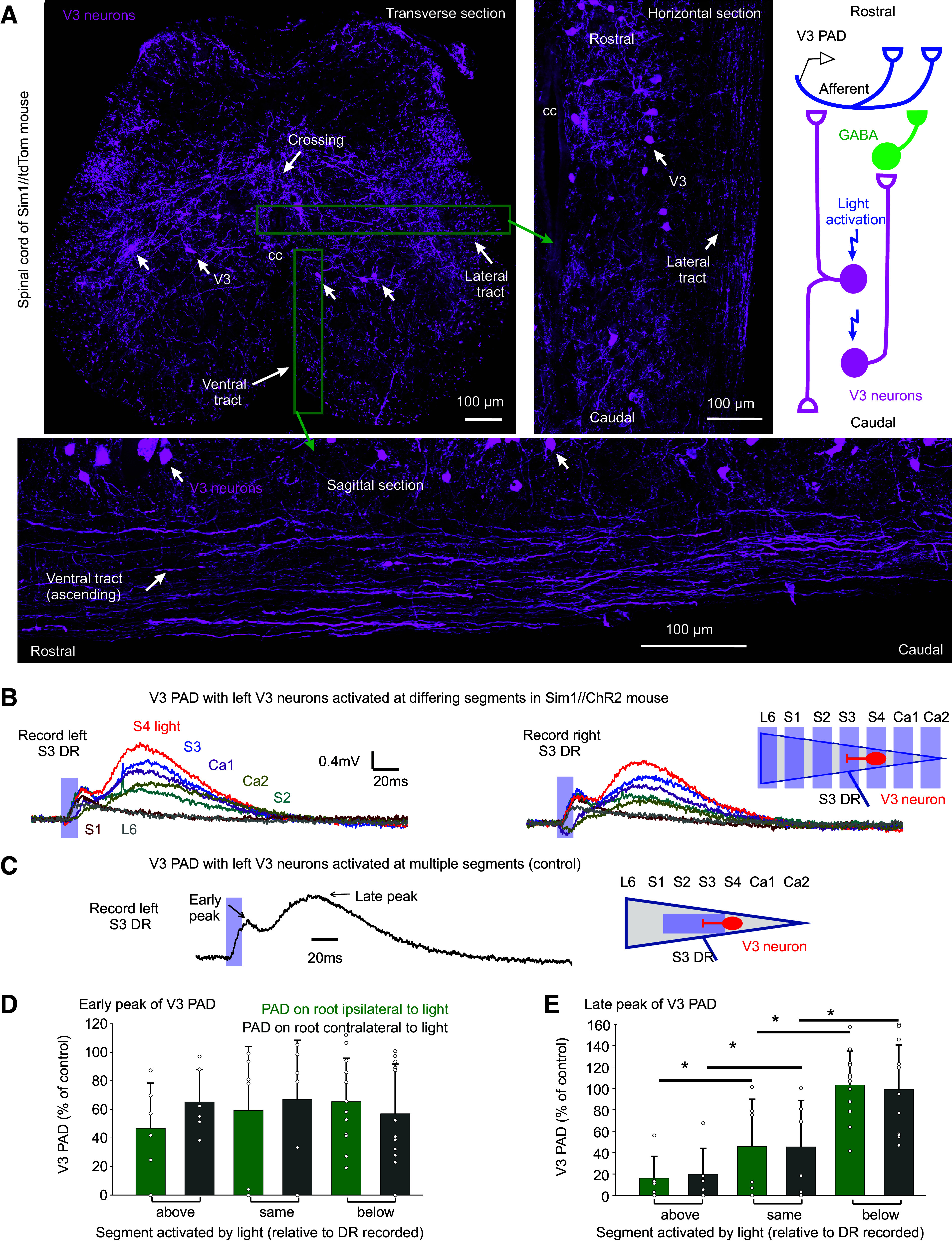
Figure 7.

Intersegmental propriospinal projections mediating PAD. A: V3 neurons in sacral S3 spinal cord of Sim1//tdTom mice shown in transverse (top left, repeated from Fig. 4A), sagittal (bottom), and horizontal (right) planes (the latter two at approximate locations indicated by green boxes), showing axonal tracts in the white matter formed by V3 neurons. Also, some V3 axons crossed the midline, as indicated. Schematic of proposed axonal projections of V3 neurons, with V3 neurons involved in GABA PAD (late peak of V3 PAD) mainly ascending from the lower sacral V3 neurons, and V3 neurons involved in glutamatergic PAD (AMPA PAD, early peak) both ascending and descending. B and C: V3 PAD in sacral S3 DRs evoked by light focused on the left lateral face of the cord in only one segment at a time (B, by turning the narrow columnated beam of Fig. 4B across the cord, as shown; 10 ms pulse, λ = 447 nm laser, 0.7 mW/mm2, 3xT). V3 PAD was similar in left and right S3 DRs (commissural). Note that S3 PAD evoked in the S3 DR by applying light at S4 (red) was largest, sometimes even larger than PAD evoked by light applied across multiple segments at and above the S3 level (C; with columnated light turned to align with cord over S2–S3, as was usual arrangement elsewhere; Fig. 4B), the latter used as our control PAD with which to normalized responses. D and E: normalized group averages of early and late peaks of V3 PAD, grouped by whether the light was applied above (rostral to), at, or below (caudal to) the segment of the root where PAD was recorded (S3 or S4 DRs). *Significant difference with applied light above or below root segmental level, P < 0.05, n = 6–12 roots per condition. DR, dorsal root; PAD, primary afferent depolarization.
