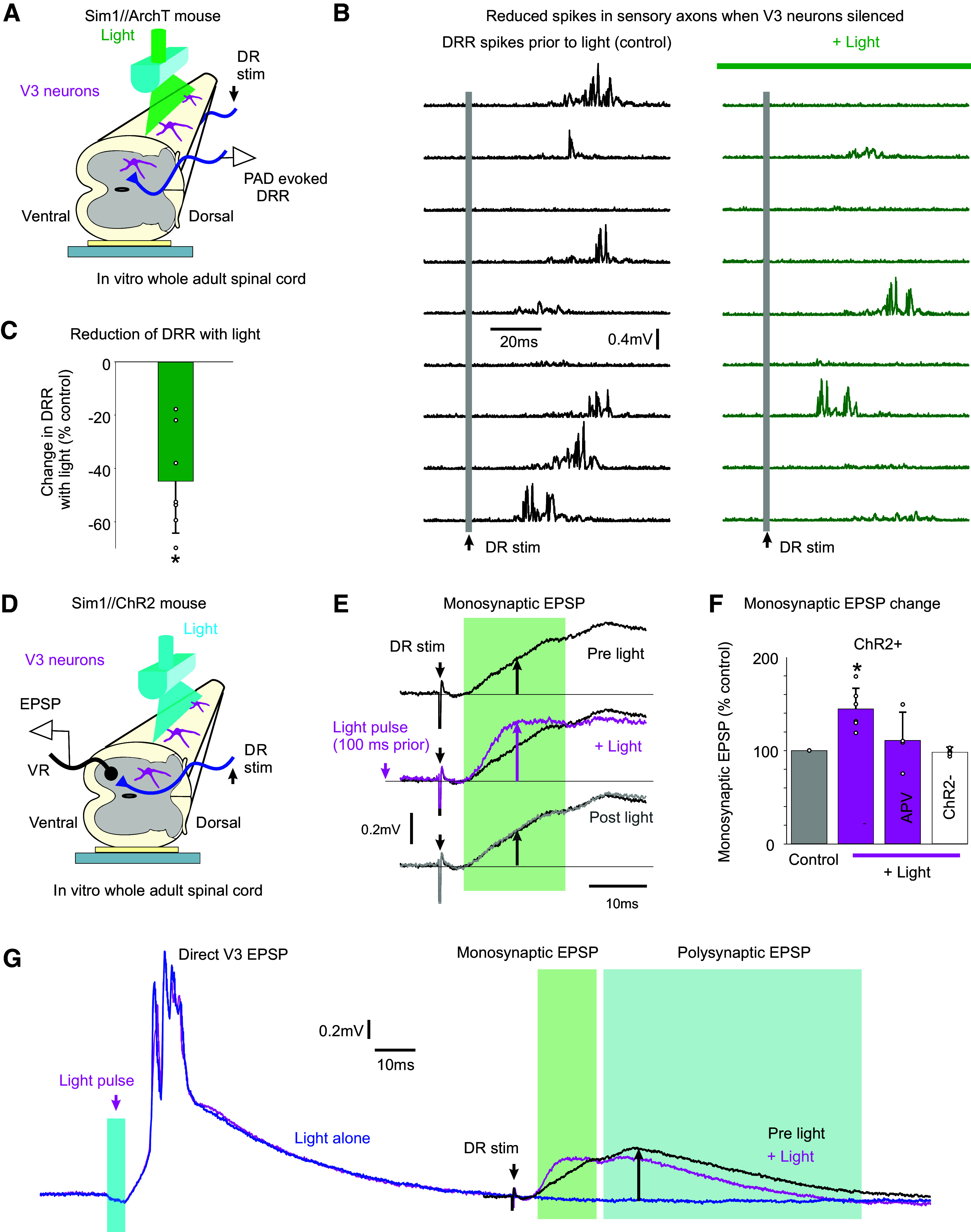
Figure 8.

V3 neurons increase spike transmission in sensory afferents. A and B: dorsal root reflex (DRR) recorded in S4 DR from stimulating the Ca1 DR (0.1 ms, 2xT; raster plot of 9 trials at 0.1 Hz) in Sim1//ArchT mice, before and after silencing V3 neurons with light (532 nm, 5 mW/mm2). The drPAD evoked by this stimulation is shown in Fig. 3A (same recording), where the rising phase of PAD produced the DRR, but here the DRR is shown in isolation by removing the slow PAD response with a 100 Hz high-pass filter and then rectifying. C: change in integrated area under rectified DRR with silencing of V3 neurons, *significant change, P < 0.05, n = 8 roots from 3 mice. D and E: motoneuron monosynaptic EPSP evoked by dorsal root stimulation (S4 DR, 0.1 ms pulse, 1.1xT) measured with grease-gap recordings from ventral root (S4 VR, giving composite EPSP of motoneuron pool; top trace in E). Light activation of the V3 neurons (10 ms pulse, λ = 447 nm laser, 0.7 mW/mm2, 3xT, laser aligned as in Fig. 4B) applied 100 ms before the same DR stimulation increased the resulting monosynaptic EPSP (magenta trace in E). F: group averages of change in monosynaptic EPSP with light, both without (as in E) and with APV (50 µM) in the bath. *Significant increase, P < 0.05, n = 6 roots without APV, n = 4 roots with APV present, from 4 mice each. G: same data as in E, but on longer time base to show direct response to the V3 activation (left) and decrease in polysynaptic reflex evoked by the DR stimulation (right, at upward arrow). PAD, primary afferent depolarization.
