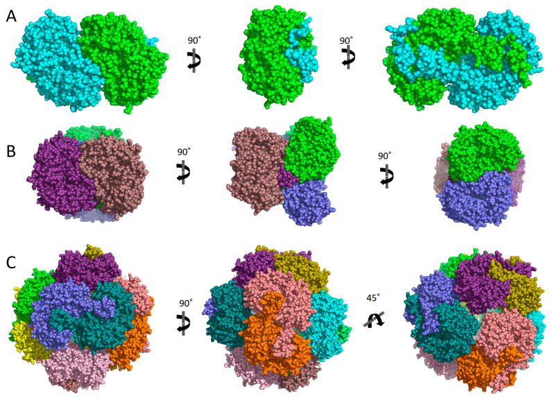
Figure 6.
Representative examples of hALDH quaternary assemblies. Each structure is shown from three different views, as indicated. Space-filling models are used, and the different colours represent different monomers. (A) hALDH3A1 dimer (PDBid: 3SZB). (B) hALDH1A1 tetramer (PDBid: 4WB9). (C) hALDH5A1 dodecamer of reduced protein (PDBid: 2W8O).

