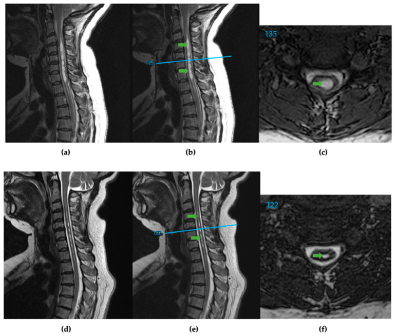
Figure 1.
(a) T2 weighted sagittal spine scan of subject 91 before surgical treatment. (b) Preoperative sagittal spine scan with cervical syringes labeled, green arrows, and the reference line, blue, for axial slice location. (c) Preoperative axial slice 135 with labeled syrinx, green. (d) T2 weighted sagittal spine scan after surgical treatment. (e) Postoperative sagittal spine scan with diminished cervical syringes labeled, green arrows, and the reference line, blue. (f) Postoperative axial slice 122 with labeled syrinx cross-section, green.

