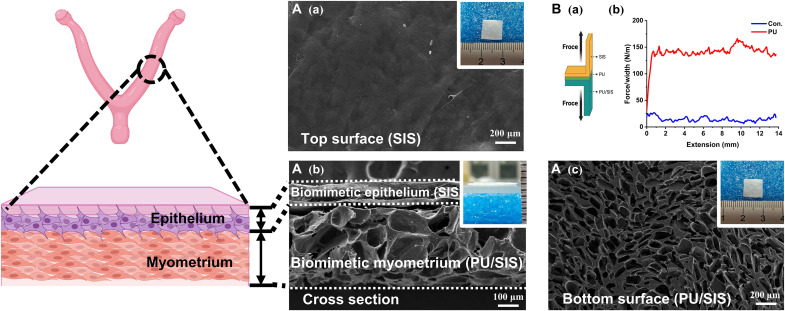
Fig. 1. Preparation of the bilayered SPS composites biomimicking the uterine structure.
(A) Scanning electron microscope (SEM) images of the top [(Aa) scale bar, 200 μm)] cross-sectional [(Ab) scale bar, 100 μm], and bottom [(Ac) scale bar, 200 μm] micromorphology of the SPS composites. Inside the white frame is the macroscopic morphology of the SPS. (B) Schematic of the 180° peel test (Ba). The force-extension curves of the bilayered SPS composites (Bb).

