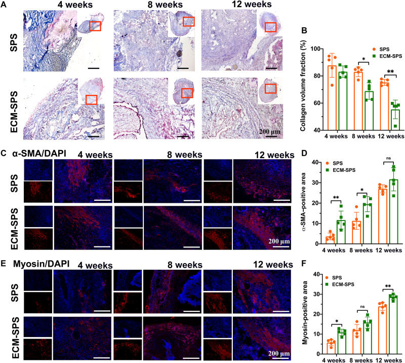
Fig. 7. The collagen remodeling and smooth muscle regeneration by various interventions.
(A) Masson’s trichrome staining of the reconstructed uterine segments. Scale bars, 200 μm. (B) Statistical analysis of collagen volume fraction on the Masson’s trichrome–stained slides (n = 5 independent samples, *P < 0.05 and **P < 0.01). (C) Immunostaining of α-SMA for myometrium regeneration in the reconstructed uterine segments. Scale bars, 200 μm. (D) Statistical analysis of α-SMA–positive area (n = 5 independent samples, *P < 0.05, **P < 0.01, and ns). (E) Immunostaining of myosin for myometrium regeneration in the reconstructed uterine segments. Scale bars, 200 μm. (F) Statistical analysis of myosin positive area (n = 5 independent samples, *P < 0.05, **P < 0.01, and ns).

