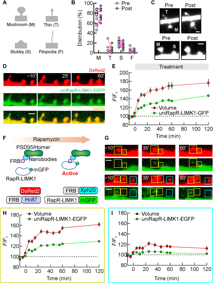
Fig. 4. Engineered LIMK1-mediated enlargement of different spine subtypes.
(A) Classification of dendritic spine types (mushroom, thin, stubby, and filopodia) based on morphological criteria. (B) A comparison of the spine type distribution in neurons expressing unimolecular rapamycin regulatable domain (uniRapR)–LIMK1 before and after rapamycin application. (C) Representative spine subtypes before and after activation by rapamycin. (D) A series of two-photon images in a dendrite of a transfected hippocampal CA1 pyramidal neuron before and after rapamycin application [green, enhanced green fluorescent protein (EGFP)–tagged uniRapR-LIMK1; red, DsRed2]. Spine volume (DsRed2, red) and amount of uniRapR-LIMK1 protein in the spine (EGFP, green) were quantified by measuring the total fluorescence intensity (F) relative to the averaged baseline fluorescence intensity (F0). (E) Spine volume and uniRapR-LIMK1 protein amount (mean ± SEM) were monitored for 120 min after rapamycin application. (F) Schematic of the strategy to accumulate engineered LIMK1 in the spines (top). The constructs used to accumulate engineered LIMK1 in the spines included nanobodies against Homer1 (Hc87) and PSD95 (Xph20) fused with FKBP12-rapamycin-binding (FRB) and RapR-LIMK1-EGFP (bottom). DsRed2 was used as a volume marker. (G) A series of two-photon images in a dendrite of CA1 pyramidal neuron transfected with (DsRed2, FRB-Xc87, FRB-Xph20, and RapR-LIMK1-EGFP) before and after rap application. Spines with rapamycin-mediated RapR-LIMK1-EGFP accumulation are denoted by yellow squares, while spines without accumulation are denoted by cerulean squares. Spine volume was monitored for 120 min after rapamycin application and quantified in spines with (H) or without (I) RapR-LIMK1 accumulation. Data are expressed as mean ± SEM. Scale bars, 1.5 μm.

