Figure 1.
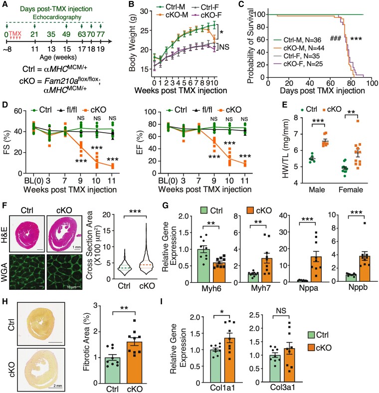
FAM210A deficiency in cardiomyocytes leads to dilated cardiomyopathy and heart failure. (A) Experimental timeline for phenotypic examination of CM-specific Fam210a cKO mice. (B) The body weight was measured weekly post-TMX-induced Fam210a KO in male and female mice. n = 11 for Ctrl and cKO male (M). n = 7 for Ctrl female (F) and n = 10 for cKO female. (C) The survival rate in male and female mice post-KO. ###P < 0.001 for M and ***P < 0.001 for F by log-rank test. (D) Fractional shortening (FS) and ejection fraction (EF) were measured by echocardiography in cKO and control mice. n = 5M + 4F for Ctrl and cKO. n = 5M + 2F for Fam210a fl/fl control mice. NS, not significant for fl/fl vs. Ctrl by two-way ANOVA with Sidak's multiple comparisons. (E) Heart weight/tibia length (HW/TL) ratio in male and female cKO mice. n = 6 and 7 for male Ctrl and cKO. n = 11 for female Ctrl and cKO. (F and H) Wheat germ agglutinin (WGA) staining for the cross-sectional area of CMs (F; n = 3 hearts with >1000 CMs quantified per heart) and picrosirius red staining of collagen deposition (H; n = 5M + 4F for Ctrl and cKO) in the hearts of control and cKO mice at ∼65 days post-KO. (G and I) Cardiac hypertrophy (G) and fibrosis (I) marker gene expression at ∼65 days post-KO (n = 5M + 4F). *P < 0.05; **P < 0.01; ***P < 0.001 by two-way ANOVA with Sidak's multiple comparisons test (B and D), Student’s t-test (E and G–I), and Mann–Whitney test (F and Nppb in G).

