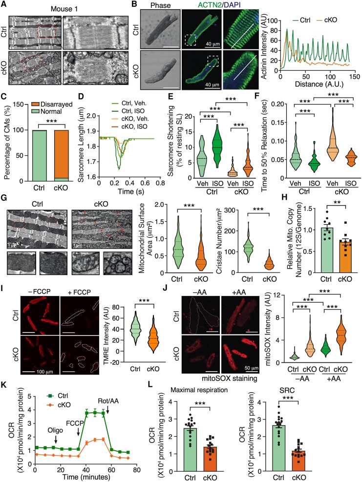
Figure 2.
Cardiomyocyte-specific deletion of Fam210a causes myofilament disarray and mitochondrial dysfunction in cardiomyocytes. (A) A representative electron microscopy image shows a disturbed sarcomeric structure in CMs from whole heart tissue sections of a late-stage Fam210a cKO heart (∼65 days post-KO). (B) Immunofluorescence staining of ACTN2 indicates myofilament disarray in isolated CMs from cKO hearts at ∼65 days post-KO. Right panels: quantification of fluorescence intensity distribution (AU, arbitrary unit) along the white line in the IF image. (C) Quantification of disarrayed CM percentile from IF staining in isolated CMs from hearts of control or cKO mice. n > 2500 CMs were quantified from four hearts. (D–F) FAM210A deficiency reduces sarcomere length and attenuates the contractility of isolated CMs from cKO hearts at ∼65 days post-KO with or without ISO stimulation. n = 82/79/77/68 CMs were quantified for Ctrl-Veh, Ctrl-ISO, cKO-Veh, and cKO-ISO from four hearts. (G) Electron microscopy shows decreased cristae and mitochondrial size in Fam210a cKO CMs from whole heart tissue sections. Triangles indicate fragmented mitochondria. The mitochondrial surface area and cristae number are quantified in >200 mitochondria from n = 3 hearts (cKO vs. Ctrl). (H) The mtDNA copy number in control and cKO hearts was measured by qPCR using primers targeting the mitochondrial 12S rDNA locus (n = 5M + 4F). qPCR of the nuclear genomic DNA was used as a normalizer. (I) Mitochondrial membrane potential (Δψm) was determined by TMRE staining in isolated CMs from late-stage Fam210a cKO hearts. For the FCCP treatment, 10 μM FCCP was added into the medium for 5 min before imaging. Greater than 600 CMs from four hearts were quantified. The TMRE intensity was normalized by the intensity after FCCP treatment. (J) The ROS level in isolated CMs from late-stage cKO hearts. Greater than 200 CMs from three hearts were quantified. AA, antimycin A (2 mM for 30 min). (K and L) The mitochondrial respiratory activity in isolated CMs from control and cKO hearts at ∼65 days post-KO was measured by the Seahorse assay. n = 16 biological replicates for Ctrl and n = 15 for cKO from CM isolations in three hearts. SRC, spare respiratory capacity (OCRMax − OCRBas). *P < 0.05, **P < 0.01, ***P < 0.001 by χ2 test (C), Kruskal–Wallis test with Dunn’s multiple comparisons test (E, F, and J), Student’s t-test (H and L), and Mann–Whitney test (G and I).

