Figure 2.

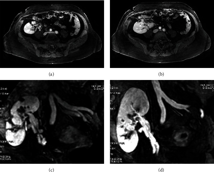
Magnetic resonance angiography (MRA) reconstructed MIP imaging showing peripheral parenchymal defects (a, b) and the permeability of the artery and the vein's graft (c, d).

Magnetic resonance angiography (MRA) reconstructed MIP imaging showing peripheral parenchymal defects (a, b) and the permeability of the artery and the vein's graft (c, d).