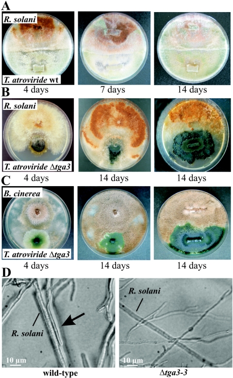
FIG. 3.
Biocontrol assays for analyzing mycoparasitic ability. (A to C) Plate confrontation assays of T. atroviride wild-type strain P1 (wt) (A) and the Δtga3-3 mutant with R. solani (B) and B. cinerea (C). In the plates on the right in panels B and C, the tga3-negative mutant was grown until it covered about one-half of the plate, and then the host fungi were inoculated. Pictures were taken 4, 7, and 14 days (A) or 4 and 14 days (B and C) after inoculation. (D) Microscopic examination of the mycoparasitic interaction between the T. atroviride wild type or the Δtga3-3 mutant and R. solani. Attachment of the wild type to the host's hyphae is indicated by the arrow.

