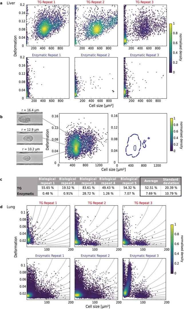
Extended Data Fig. 4. Physical phenotype characterisation of cells isolated mechanically and enzymatically from murine lung and liver.
a, Scatter plots of deformation vs cell size for cells isolated from mouse liver tissue using a tissue grinder (TG) or enzymatic dissociation, showing the enrichment of hepatocytes following mechanical dissociation for 3 independent biological repeats. b, Scatter plot of deformation vs cell size showing 3 clusters of cells that correspond to hepatocytes of different sizes; with the corresponding kernel density estimate (KDE) plot and representative images (r = radius of cells). c, Percentage of hepatocytes to the total number of liver cells, as detected by RT-DC for five independent biological repeats. d, Scatter plots of deformation vs cell size for cells isolated from mouse lung tissue using a tissue grinder or enzymatic dissociation for 3 independent biological repeats.

