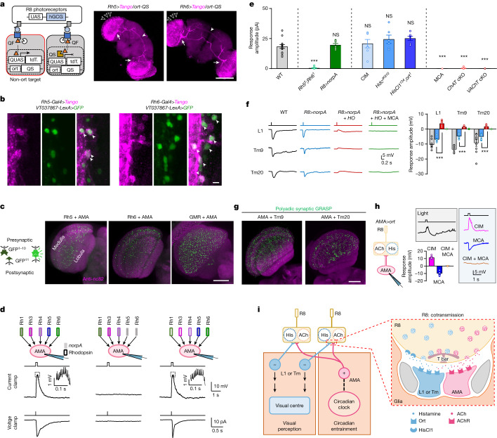
Fig. 3. ACh and histamine act on distinct neurons.
a, ort-independent postsynaptic neurons. Left: schematic of trans-Tango tracing with ort-QS excluding QF-driven tdTomato (tdT.) expression in ort-expressing neurons; right: ort-independent postsynaptic neurons of pR8 and yR8 photoreceptors. Open arrowheads mark multicolumnar arborization, arrows mark accessory medulla innervation, and filled arrowheads mark the arcuate commissure. Scale bar, 100 μm. b, VT037867-labelled neurons overlap with ort-independent postsynaptic neurons of pR8 (left) and yR8 (right) photoreceptors. Arrowheads mark co-labelled cells. Scale bar, 5 μm. c, Connections between R8 photoreceptors and AMA neurons. Left: schematic of GRASP labelling; right: GRASP between AMA neurons and pR8 (Rh5-Gal4), yR8 (Rh6-Gal4) or all (GMR-Gal4) photoreceptors. Scale bar, 50 μm. d, AMA neurons are excited by R8 photoreceptors. Top: schematic of photoreceptor manipulation: wild-type flies (left), Rh52;Rh61 flies (middle) and norpAP41 flies with norpA rescued in R8 photoreceptors (right). Middle: representative light-induced depolarization; insets represent spikes outlined by the dashed line. Bottom: representative light-induced inward current. Light: 470 nm, 5.56 × 10⁷ photons μm−2 s−1, 200 ms (current clamp) and 2 ms (voltage clamp). e, Light-induced saturated responses in AMA neurons. cKO, conditional knockout. f, Light-induced hyperpolarization of ort-expressing postsynaptic neurons of R8 photoreceptors. Left: representative light-induced hyperpolarization of L1, Tm9 and Tm20 neurons. Right: pooled saturated response amplitudes. g, AMA and Tm9 or Tm20 neurons share the same polyadic R8 synapse. h, Histamine-mediated responses in ort-expressing AMA neurons. Left: schematic of R8 inputs. Right: representative biphasic light responses in normal saline (top left); depolarization in CIM, hyperpolarization in MCA and no response in CIM + MCA (right) and pooled data (bottom left). i, A model of postsynaptic segregation of cotransmission. Although histamine signalling dominates in transmission from R8 photoreceptors to L1 and Tm pathways, very minor ACh signalling also exists in this pathway (f). Light in d–f,h: 470 nm, 5.56 × 10⁷ photons μm−2 s−1, duration of 2 ms (e,f) and 200 ms (d,h). Pooled data are shown as mean ± s.e.m. ***P < 0.001; NS, not significant. Statistical analysis is summarized in Supplementary Table 2.

