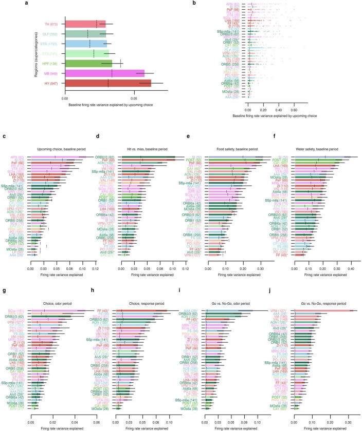
Extended Data Fig. 5. Regional distribution of regressor information.
a, Average variance explained per-cell by upcoming choice at baseline (1 s pre-odor average) for high-level brain regions. CTXsp, cortical subplate; HB, hindbrain; OLF, olfactory areas; STR, striatum; TH, thalamus; HPF, hippocampal formation; MB, midbrain; HY, hypothalamus. b, Variance explained per-cell by upcoming choice at baseline; each dot represents the variance explained for a single cell in a given region. c–j, Average variance explained per-cell for regressors described in Extended Data Fig. 3c. For all panels, bars are averages across neurons within a given region; black lines give the 95% distribution of information per cell in a region. Numbers in parentheses are unit counts per region. Color codes are according to the Allen Institute Mouse Brain Atlas colormap. Dashed lines indicate the circular permutation null distribution for a given regressor and region. Regions are ordered by variance explained. Note that Fig. 2e shows a subset (only regions with average variance explained significantly greater than the null) of data shown in panel c here.

