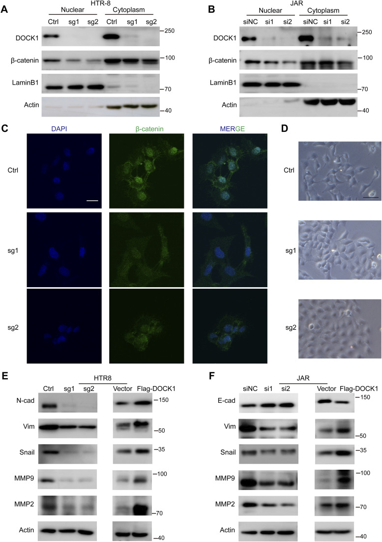
Figure 4. DOCK1 promotes the epithelial–mesenchymal transition (EMT) process.
(A) β-catenin protein levels in different subcellar locations of HTR-8 cells depleted of DOCK1 analyzed by Western blot. (B) Western blot analysis of β-catenin protein levels in different subcellar locations of JAR cells transfected with siDOCK1 or siNC. (C) Representative images of β-catenin immunofluorescence staining in DOCK1 knockout HTR-8 cells. Scale bar: 25 μm. (D) Morphological changes in HTR-8 cells after DOCK1 knockout. Scale bar: 50 μm. (E) Western blot results showing EMT or metastasis-related protein levels in HTR-8 cells after DOCK1 knockout or overexpression. (F) Western blot results showing EMT or metastasis-related protein levels in JAR cells with silenced or overexpressed DOCK1.

