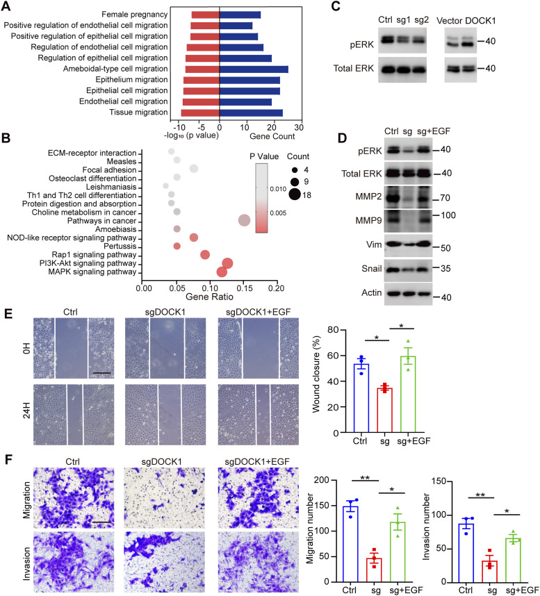
Figure 5. DOCK1 promotes cell motility via the ERK signaling pathway.
(A) Gene ontology (GO) analysis showing the significant relevance of the top 10 most prominent terms associated with differentially expressed genes (DEGs) after DOCK1 knockout in HTR-8 cells. (B) Kyoto Encyclopedia of Genes and Genomes pathway analysis identified the top 15 enriched pathways associated with DEGs. Functional enrichment was used to identify the biological processes associated with the DEGs after DOCK1 knockout in HTR-8 cells. (C) Phosphorylation levels of ERK protein in HTR-8 cells after DOCK1 repression or overexpression. (D) Metastasis-related protein levels after activation of the ERK signaling pathway in DOCK1 knockout HTR-8 cells. (E) Wound healing assay results showing migration ability of HTR-8 cells with DOCK1 knockout after EGF treatment activated the ERK signaling pathway (left). The rate of wound closure was quantified (right). Scale bar: 200 μm. (F) Transwell assays results showing the migration and invasion of HTR-8 cells with DOCK1 knockout after EGF treatment (left). The number of migrating and invading cells was quantified (right). Scale bar: 100 μm. In (E, F), each group, n = 3 biological replicates (*P < 0.05; unpaired two-tailed t test).

