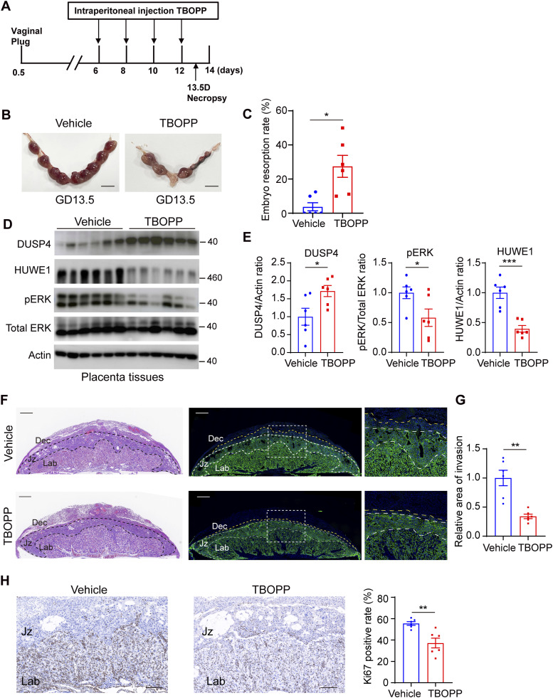
Figure 9. DOCK1 deficiency triggers embryo miscarriage in pregnant mice models.
(A) Schematic illustration of the protocol for inhibiting DOCK1 during murine pregnancy. Experimental mice received vehicle control or TBOPP on GDs 6, 8, 10, and 12 through intraperitoneal injections, and were euthanatized on GDs 13.5. (B) Morphology of pregnant uteri with embryo implantation sites on GD 13.5 in mouse models after vehicle control or TBOPP treatments (n = 6 mice for each group). Scale bar: 1 cm. (C) Embryo resorption rate was calculated. (D) Western blot assays were performed to detect the levels of DUSP4, phosphorylated ERK, and HUWE1 after TBOPP treatment. (E) Quantification of DUSP4, phosphorylated ERK, and HUWE1 protein levels. (F) Representative HE-stained images of placentas and CK7 immunofluorescence of trophoblast cell invasion into the decidua after TBOPP treatment (left). Based on the HE and CK7 staining, distinct layers—Dec (decidua), Jz (junctional zone), and Lab (labyrinth)—are delineated by black lines in HE images and white lines in immunofluorescence images. Boxes on the left correspond to magnified sections on the right, with the region between the yellow and white lines indicating the trophoblast cell invasion area into the decidua. Scale bar: 500 μm. (G) The area of trophoblast cell invasion into the decidua was quantified. (H) Representative images of Ki67 immunostaining in placental tissues after TBOPP treatment (left). Percentage of Ki67-positive cells was calculated (right). Scale bar: 200 μm. In (C, E, G, H), each group, n = 6 mice (*P < 0.05; **P < 0.01; ***P < 0.001, unpaired two-tailed t test).

